File list
This special page shows all uploaded files.
| Date | Name | Thumbnail | Size | User | Description | Versions |
|---|---|---|---|---|---|---|
| 14:52, 8 August 2013 | Test.mp3 (file) | 5.65 MB | Seung Park | Testing out Html5mediator | 1 | |
| 17:19, 8 August 2013 | Test.mp4 (file) | 4.17 MB | Seung Park | More testing! | 2 | |
| 14:25, 14 August 2013 | Test.jpg (file) |  |
270 KB | Peter Anderson | This is a test of the uploading system. | 1 |
| 13:49, 15 August 2013 | IPLab1MyocardialInfarction1.jpg (file) | 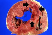 |
38 KB | Seung Park | 1 | |
| 13:49, 15 August 2013 | IPLab1MyocardialInfarction2.jpg (file) | 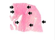 |
22 KB | Seung Park | 1 | |
| 13:49, 15 August 2013 | IPLab1MyocardialInfarction3.jpg (file) | 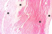 |
65 KB | Seung Park | 1 | |
| 13:50, 15 August 2013 | IPLab1MyocardialInfarction4.jpg (file) | 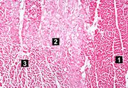 |
85 KB | Seung Park | 1 | |
| 13:50, 15 August 2013 | IPLab1MyocardialInfarction5.jpg (file) |  |
77 KB | Seung Park | 1 | |
| 13:50, 15 August 2013 | IPLab1MyocardialInfarction6.jpg (file) |  |
60 KB | Seung Park | 1 | |
| 13:50, 15 August 2013 | IPLab1MyocardialInfarction7.jpg (file) |  |
60 KB | Seung Park | 1 | |
| 15:12, 15 August 2013 | IPLab1KidneyInfarction1.jpg (file) |  |
70 KB | Seung Park | 1 | |
| 15:12, 15 August 2013 | IPLab1KidneyInfarction2.jpg (file) | 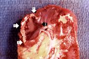 |
47 KB | Seung Park | 1 | |
| 15:12, 15 August 2013 | IPLab1KidneyInfarction3.jpg (file) |  |
11 KB | Seung Park | 1 | |
| 15:12, 15 August 2013 | IPLab1KidneyInfarction4.jpg (file) | 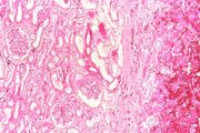 |
71 KB | Seung Park | 1 | |
| 15:12, 15 August 2013 | IPLab1KidneyInfarction5.jpg (file) | 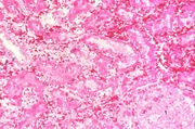 |
64 KB | Seung Park | 1 | |
| 15:12, 15 August 2013 | IPLab1KidneyInfarction6.jpg (file) | 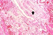 |
64 KB | Seung Park | 1 | |
| 15:12, 15 August 2013 | IPLab1KidneyInfarction7.jpg (file) | 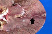 |
43 KB | Seung Park | 1 | |
| 16:14, 15 August 2013 | IPLab1LungAbscess1.jpg (file) | 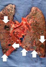 |
30 KB | Seung Park | 1 | |
| 16:15, 15 August 2013 | IPLab1LungAbscess2.jpg (file) | 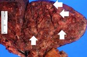 |
57 KB | Seung Park | 1 | |
| 16:15, 15 August 2013 | IPLab1LungAbscess3.jpg (file) | 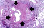 |
62 KB | Seung Park | 1 | |
| 16:15, 15 August 2013 | IPLab1LungAbscess4.jpg (file) | 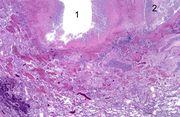 |
75 KB | Seung Park | 1 | |
| 16:15, 15 August 2013 | IPLab1LungAbscess5.jpg (file) | 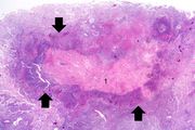 |
56 KB | Seung Park | 1 | |
| 16:15, 15 August 2013 | IPLab1LungAbscess6.jpg (file) | 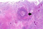 |
66 KB | Seung Park | 1 | |
| 16:15, 15 August 2013 | IPLab1LungAbscess7.jpg (file) | 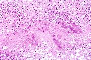 |
79 KB | Seung Park | 1 | |
| 16:15, 15 August 2013 | IPLab1LungAbscess8.jpg (file) | 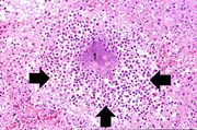 |
85 KB | Seung Park | 1 | |
| 01:17, 16 August 2013 | IPLab1FatNecrosis1.jpg (file) | 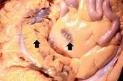 |
36 KB | Seung Park | 1 | |
| 01:17, 16 August 2013 | IPLab1FatNecrosis2.jpg (file) | 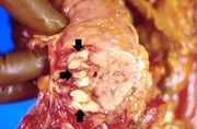 |
42 KB | Seung Park | 1 | |
| 01:18, 16 August 2013 | IPLab1FatNecrosis3.jpg (file) |  |
65 KB | Seung Park | 1 | |
| 01:18, 16 August 2013 | IPLab1FatNecrosis4.jpg (file) |  |
76 KB | Seung Park | 1 | |
| 01:18, 16 August 2013 | IPLab1FatNecrosis5.jpg (file) |  |
68 KB | Seung Park | 1 | |
| 01:18, 16 August 2013 | IPLab1FatNecrosis6.jpg (file) |  |
83 KB | Seung Park | 1 | |
| 01:18, 16 August 2013 | IPLab1FatNecrosis7.jpg (file) | 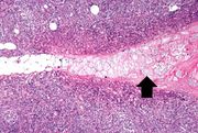 |
81 KB | Seung Park | 1 | |
| 01:18, 16 August 2013 | IPLab1FatNecrosis8.jpg (file) | 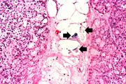 |
72 KB | Seung Park | 1 | |
| 01:19, 16 August 2013 | IPLab1FatNecrosis9.jpg (file) | 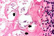 |
52 KB | Seung Park | 1 | |
| 02:49, 16 August 2013 | IPLab1Tuberculosis1.jpg (file) | 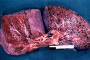 |
66 KB | Seung Park | 1 | |
| 02:49, 16 August 2013 | IPLab1Tuberculosis2.jpg (file) | 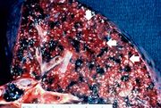 |
72 KB | Seung Park | 1 | |
| 02:49, 16 August 2013 | IPLab1Tuberculosis3.jpg (file) | 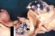 |
42 KB | Seung Park | 1 | |
| 02:50, 16 August 2013 | IPLab1Tuberculosis4.jpg (file) |  |
21 KB | Seung Park | 1 | |
| 02:50, 16 August 2013 | IPLab1Tuberculosis5.jpg (file) | 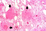 |
58 KB | Seung Park | 1 | |
| 02:50, 16 August 2013 | IPLab1Tuberculosis6.jpg (file) | 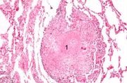 |
64 KB | Seung Park | 1 | |
| 02:50, 16 August 2013 | IPLab1Tuberculosis7.jpg (file) | 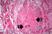 |
64 KB | Seung Park | 1 | |
| 03:40, 16 August 2013 | IPLab1Prostate1.jpg (file) | 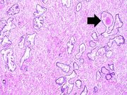 |
86 KB | Seung Park | 1 | |
| 03:40, 16 August 2013 | IPLab1Prostate2.jpg (file) | 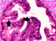 |
61 KB | Seung Park | 1 | |
| 03:41, 16 August 2013 | IPLab1Prostate3.jpg (file) | 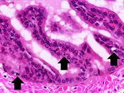 |
60 KB | Seung Park | Another high-power photomicrograph of the prostatic epithelium shows cells with pyknotic and fragmented nuclei (arrows). Note that the cytoplasm is condensed and hypereosinophilic. | 1 |
| 03:41, 16 August 2013 | IPLab1Prostate4.jpg (file) | 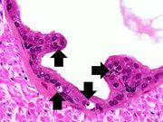 |
46 KB | Seung Park | Still another high-power photomicrograph of the prostatic epithelium demonstrates cells with pyknotic and fragmented nuclei (arrows). Again note the condensed and hypereosinophilic cytoplasm. | 1 |
| 03:42, 16 August 2013 | IPLab1Prostate5.jpg (file) | 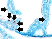 |
35 KB | Seung Park | This photomicrograph of prostatic epithelium demonstrates an in situ immunohistochemical technique that is used to identify the DNA fragments characteristic of apoptotic nuclei. This technique, terminal deoxynucleotidyl transferase-mediated dUTP-biotin... | 1 |
| 03:42, 16 August 2013 | IPLab1Prostate6.jpg (file) | 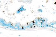 |
27 KB | Seung Park | This is a higher-power photomicrograph of prostatic epithelium with the TUNEL staining. Note the apoptotic cells (brown nuclei) in the epithelium as well as those floating freely. | 1 |
| 23:29, 18 August 2013 | IPLab2Hypertrophy1.jpg (file) | 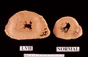 |
30 KB | Seung Park | This is a gross photograph of a cross section of a normal human heart taken at autopsy (right) and the heart from this case, which demonstrates concentric hypertrophy of the left ventricular wall. Note the marked thickening of the left ventricular wall... | 1 |
| 23:29, 18 August 2013 | IPLab2Hypertrophy5.jpg (file) | 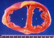 |
47 KB | Seung Park | This autopsy specimen was taken from another patient who had cardiac hypertrophy and congestive heart failure that resulted in dilation of the cardiac chambers. This heart was markedly enlarged (700 grams) but the congestive failure leads to dilation o... | 1 |
| 23:30, 18 August 2013 | IPLab2Hypertrophy6.jpg (file) | 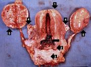 |
63 KB | Seung Park | This gross photograph shows an example of normal physiologic hypertrophy. The organs shown are an open uterus (1), cervix (2) and vagina (3), both ovaries (4) and both kidneys (5) from a woman who died shortly after normal delivery from causes unrelate... | 1 |