Difference between revisions of "File:IPLab9ARF6.jpg"
Seung Park (talk | contribs) (This high-power photomicrograph of myocardium shows the cellular detail of another Aschoff body. In this case there appears to be a multinucleated Aschoff giant cell (arrow).) |
(No difference)
|
Latest revision as of 03:53, 21 August 2013
This high-power photomicrograph of myocardium shows the cellular detail of another Aschoff body. In this case there appears to be a multinucleated Aschoff giant cell (arrow).
File history
Click on a date/time to view the file as it appeared at that time.
| Date/Time | Thumbnail | Dimensions | User | Comment | |
|---|---|---|---|---|---|
| current | 03:53, 21 August 2013 | 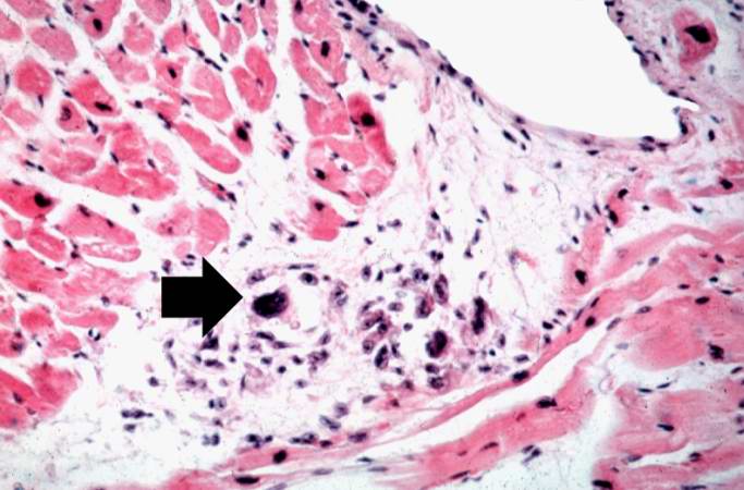 | 683 × 450 (50 KB) | Seung Park (talk | contribs) | This high-power photomicrograph of myocardium shows the cellular detail of another Aschoff body. In this case there appears to be a multinucleated Aschoff giant cell (arrow). |
- You cannot overwrite this file.
File usage
The following page links to this file: