Difference between revisions of "File:IPLab10Histo3.jpg"
Seung Park (talk | contribs) (This is an even higher-power photomicrograph of an area of necrosis (arrows). There is loss of cellular detail within this area. There are inflammatory cells present; however, it is difficult to differentiate the inflammatory cells from the native lymp...) |
(No difference)
|
Latest revision as of 04:06, 21 August 2013
This is an even higher-power photomicrograph of an area of necrosis (arrows). There is loss of cellular detail within this area. There are inflammatory cells present; however, it is difficult to differentiate the inflammatory cells from the native lymphocytes of the adrenal gland--which is a lymph node.
File history
Click on a date/time to view the file as it appeared at that time.
| Date/Time | Thumbnail | Dimensions | User | Comment | |
|---|---|---|---|---|---|
| current | 04:06, 21 August 2013 | 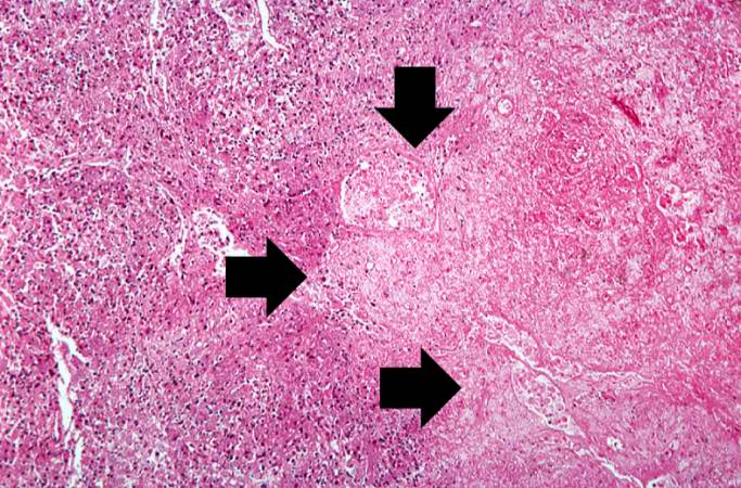 | 683 × 450 (90 KB) | Seung Park (talk | contribs) | This is an even higher-power photomicrograph of an area of necrosis (arrows). There is loss of cellular detail within this area. There are inflammatory cells present; however, it is difficult to differentiate the inflammatory cells from the native lymp... |
- You cannot overwrite this file.
File usage
The following page links to this file: