Difference between revisions of "File:IPLab10Blasto4.jpg"
Seung Park (talk | contribs) (This is a high-power photomicrograph of lung section with pleura. The pleura (1) is thickened and contains inflammatory cells and fibrin. The areas of consolidation (2) are dense and filled with inflammatory cells.) |
(No difference)
|
Latest revision as of 04:15, 21 August 2013
This is a high-power photomicrograph of lung section with pleura. The pleura (1) is thickened and contains inflammatory cells and fibrin. The areas of consolidation (2) are dense and filled with inflammatory cells.
File history
Click on a date/time to view the file as it appeared at that time.
| Date/Time | Thumbnail | Dimensions | User | Comment | |
|---|---|---|---|---|---|
| current | 04:15, 21 August 2013 | 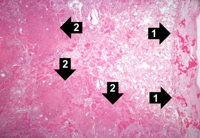 | 655 × 450 (56 KB) | Seung Park (talk | contribs) | This is a high-power photomicrograph of lung section with pleura. The pleura (1) is thickened and contains inflammatory cells and fibrin. The areas of consolidation (2) are dense and filled with inflammatory cells. |
- You cannot overwrite this file.
File usage
The following page links to this file: