Difference between revisions of "File:IPLab10Mucor4.jpg"
Seung Park (talk | contribs) (This is a higher-power photomicrograph of just the wall of the carotid artery. Note the ribbon-like clear structure with roughly parallel walls (non-septate hyphae) and right-angle branching (arrow). This is the Mucor organism.) |
(No difference)
|
Latest revision as of 04:19, 21 August 2013
This is a higher-power photomicrograph of just the wall of the carotid artery. Note the ribbon-like clear structure with roughly parallel walls (non-septate hyphae) and right-angle branching (arrow). This is the Mucor organism.
File history
Click on a date/time to view the file as it appeared at that time.
| Date/Time | Thumbnail | Dimensions | User | Comment | |
|---|---|---|---|---|---|
| current | 04:19, 21 August 2013 | 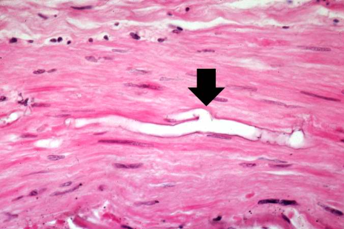 | 676 × 450 (40 KB) | Seung Park (talk | contribs) | This is a higher-power photomicrograph of just the wall of the carotid artery. Note the ribbon-like clear structure with roughly parallel walls (non-septate hyphae) and right-angle branching (arrow). This is the Mucor organism. |
- You cannot overwrite this file.
File usage
The following page links to this file: