Difference between revisions of "File:IPLab11Leishmaniasis3.jpg"
Seung Park (talk | contribs) (This is a higher-power photomicrograph of this biopsy. The ulcerated surface is seen on the top of the section. Again, note that the specimen is heavily infiltrated with inflammatory cells.) |
(No difference)
|
Latest revision as of 04:58, 21 August 2013
This is a higher-power photomicrograph of this biopsy. The ulcerated surface is seen on the top of the section. Again, note that the specimen is heavily infiltrated with inflammatory cells.
File history
Click on a date/time to view the file as it appeared at that time.
| Date/Time | Thumbnail | Dimensions | User | Comment | |
|---|---|---|---|---|---|
| current | 04:58, 21 August 2013 | 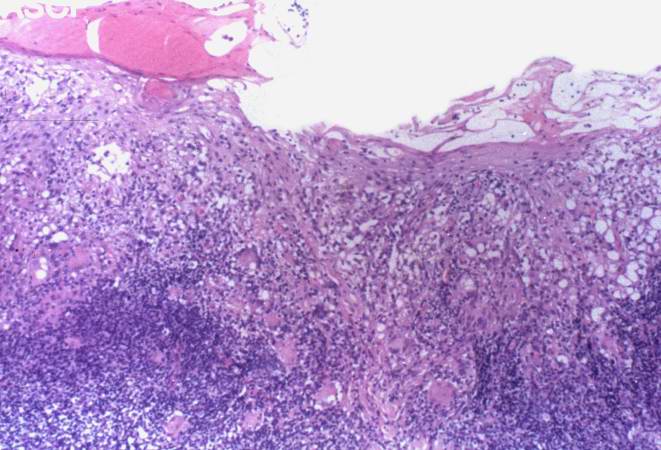 | 661 × 450 (58 KB) | Seung Park (talk | contribs) | This is a higher-power photomicrograph of this biopsy. The ulcerated surface is seen on the top of the section. Again, note that the specimen is heavily infiltrated with inflammatory cells. |
- You cannot overwrite this file.
File usage
The following page links to this file: