Difference between revisions of "File:IPLab11Ascariasis6.jpg"
Seung Park (talk | contribs) (This high-power photomicrograph of the fecal specimen from this patient shows a Giardia lamblia trophozoite. Note the two nuclei and the tapered end (that goes out of the plane of focus) containing flagella (arrow).) |
(No difference)
|
Latest revision as of 05:01, 21 August 2013
This high-power photomicrograph of the fecal specimen from this patient shows a Giardia lamblia trophozoite. Note the two nuclei and the tapered end (that goes out of the plane of focus) containing flagella (arrow).
File history
Click on a date/time to view the file as it appeared at that time.
| Date/Time | Thumbnail | Dimensions | User | Comment | |
|---|---|---|---|---|---|
| current | 05:01, 21 August 2013 | 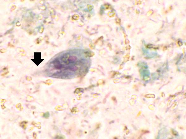 | 601 × 450 (29 KB) | Seung Park (talk | contribs) | This high-power photomicrograph of the fecal specimen from this patient shows a Giardia lamblia trophozoite. Note the two nuclei and the tapered end (that goes out of the plane of focus) containing flagella (arrow). |
- You cannot overwrite this file.
File usage
The following page links to this file: