Difference between revisions of "File:IPLab12Mesothelioma8.jpg"
Seung Park (talk | contribs) (This high-power photomicrograph shows brown asbestos bodies (1), accumulations of anthracotic pigment (2), and tumor cells (3) all adjacent to a blood vessel (4).) |
(No difference)
|
Latest revision as of 05:33, 21 August 2013
This high-power photomicrograph shows brown asbestos bodies (1), accumulations of anthracotic pigment (2), and tumor cells (3) all adjacent to a blood vessel (4).
Anthracotic pigment is coal dust deposited in the lungs--it is seen in coal miners, city-dwellers, and smokers.
File history
Click on a date/time to view the file as it appeared at that time.
| Date/Time | Thumbnail | Dimensions | User | Comment | |
|---|---|---|---|---|---|
| current | 05:33, 21 August 2013 | 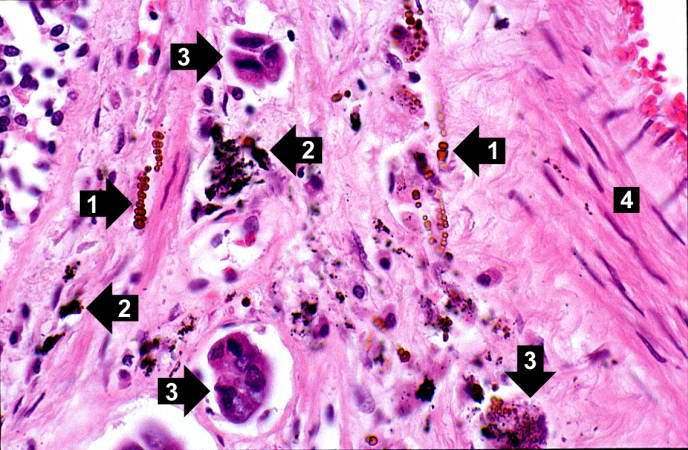 | 688 × 450 (68 KB) | Seung Park (talk | contribs) | This high-power photomicrograph shows brown asbestos bodies (1), accumulations of anthracotic pigment (2), and tumor cells (3) all adjacent to a blood vessel (4). |
- You cannot overwrite this file.
File usage
The following page links to this file: