Difference between revisions of "File:IPLab12Mesothelioma11.jpg"
Seung Park (talk | contribs) (Scanning electron micrograph of asbestos bodies. Note the rough surface and the beaded appearance caused by the material adhering to the surface of the asbestos fiber.) |
(No difference)
|
Latest revision as of 05:34, 21 August 2013
Scanning electron micrograph of asbestos bodies. Note the rough surface and the beaded appearance caused by the material adhering to the surface of the asbestos fiber.
File history
Click on a date/time to view the file as it appeared at that time.
| Date/Time | Thumbnail | Dimensions | User | Comment | |
|---|---|---|---|---|---|
| current | 05:34, 21 August 2013 | 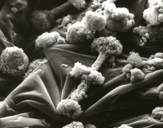 | 571 × 450 (35 KB) | Seung Park (talk | contribs) | Scanning electron micrograph of asbestos bodies. Note the rough surface and the beaded appearance caused by the material adhering to the surface of the asbestos fiber. |
- You cannot overwrite this file.
File usage
The following page links to this file: