Difference between revisions of "File:IPLab13Myelomeningocele1.jpg"
Seung Park (talk | contribs) (This is a gross photograph of the fetus at autopsy. Note the defect in the lower lumbar region of the spinal column (arrow). The myelomeningocele can be seen protruding from this defect.) |
(No difference)
|
Latest revision as of 05:44, 21 August 2013
This is a gross photograph of the fetus at autopsy. Note the defect in the lower lumbar region of the spinal column (arrow). The myelomeningocele can be seen protruding from this defect.
A myelomeningocele is the herniation of the spinal cord--within the meninges--through a defect in the vertebral canal.
File history
Click on a date/time to view the file as it appeared at that time.
| Date/Time | Thumbnail | Dimensions | User | Comment | |
|---|---|---|---|---|---|
| current | 05:44, 21 August 2013 | 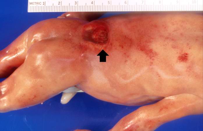 | 697 × 450 (27 KB) | Seung Park (talk | contribs) | This is a gross photograph of the fetus at autopsy. Note the defect in the lower lumbar region of the spinal column (arrow). The myelomeningocele can be seen protruding from this defect. |
- You cannot overwrite this file.
File usage
The following page links to this file: