Difference between revisions of "File:IPLab13BiliaryAtresia2.jpg"
Seung Park (talk | contribs) (This medium-power photomicrograph of liver shows an area of portal fibrosis and bile duct proliferation (arrows). Adjacent to this fibrotic portal region, hepatocytes are seen separated by dilated sinusoids. Throughout this section are found accumulati...) |
(No difference)
|
Latest revision as of 05:47, 21 August 2013
This medium-power photomicrograph of liver shows an area of portal fibrosis and bile duct proliferation (arrows). Adjacent to this fibrotic portal region, hepatocytes are seen separated by dilated sinusoids. Throughout this section are found accumulations of yellow-brown bile pigment.
File history
Click on a date/time to view the file as it appeared at that time.
| Date/Time | Thumbnail | Dimensions | User | Comment | |
|---|---|---|---|---|---|
| current | 05:47, 21 August 2013 | 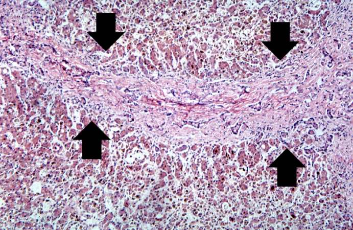 | 691 × 450 (95 KB) | Seung Park (talk | contribs) | This medium-power photomicrograph of liver shows an area of portal fibrosis and bile duct proliferation (arrows). Adjacent to this fibrotic portal region, hepatocytes are seen separated by dilated sinusoids. Throughout this section are found accumulati... |
- You cannot overwrite this file.
File usage
The following page links to this file: