Difference between revisions of "File:IPLab13Hyaline7.jpg"
Seung Park (talk | contribs) (This high-power photomicrograph shows an airway with adjacent lung tissue. Some alveoli have hyaline membranes (arrows). There is severe congestion of the interstitium throughout this section.) |
(No difference)
|
Latest revision as of 05:51, 21 August 2013
This high-power photomicrograph shows an airway with adjacent lung tissue. Some alveoli have hyaline membranes (arrows). There is severe congestion of the interstitium throughout this section.
File history
Click on a date/time to view the file as it appeared at that time.
| Date/Time | Thumbnail | Dimensions | User | Comment | |
|---|---|---|---|---|---|
| current | 05:51, 21 August 2013 | 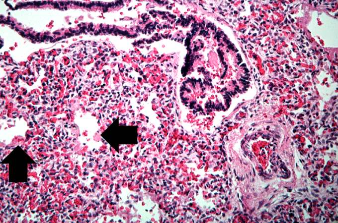 | 680 × 450 (93 KB) | Seung Park (talk | contribs) | This high-power photomicrograph shows an airway with adjacent lung tissue. Some alveoli have hyaline membranes (arrows). There is severe congestion of the interstitium throughout this section. |
- You cannot overwrite this file.
File usage
The following page links to this file: