Difference between revisions of "File:IPLab13WT4.jpg"
Seung Park (talk | contribs) (This lowest-power view shows the tumor itself; no tissue is present that can be readily identified as normal kidney. There does appear to be a capsule surrounding the tumor. Eosinophilic bands are seen surrounding basophilic islands of cells. These cor...) |
(No difference)
|
Latest revision as of 05:55, 21 August 2013
This lowest-power view shows the tumor itself; no tissue is present that can be readily identified as normal kidney. There does appear to be a capsule surrounding the tumor. Eosinophilic bands are seen surrounding basophilic islands of cells. These correspond to the two types of tissue in this tumor--the basophilic cellular component termed "blastema" can be distinguished from less cellular eosinophilic areas.
File history
Click on a date/time to view the file as it appeared at that time.
| Date/Time | Thumbnail | Dimensions | User | Comment | |
|---|---|---|---|---|---|
| current | 05:55, 21 August 2013 | 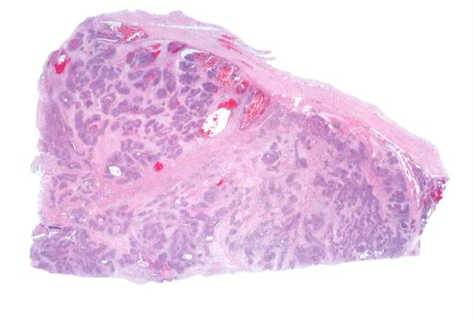 | 665 × 450 (30 KB) | Seung Park (talk | contribs) | This lowest-power view shows the tumor itself; no tissue is present that can be readily identified as normal kidney. There does appear to be a capsule surrounding the tumor. Eosinophilic bands are seen surrounding basophilic islands of cells. These cor... |
- You cannot overwrite this file.
File usage
The following page links to this file: