Difference between revisions of "File:IPLab13WT5.jpg"
Seung Park (talk | contribs) (This low-power photomicrograph of tumor shows the two cell types making up this neoplasm. The basophilic cellular component termed "blastema" (1) can be distinguished from less cellular eosinophilic areas with fibroblast-like cells (2).) |
(No difference)
|
Latest revision as of 05:55, 21 August 2013
This low-power photomicrograph of tumor shows the two cell types making up this neoplasm. The basophilic cellular component termed "blastema" (1) can be distinguished from less cellular eosinophilic areas with fibroblast-like cells (2).
File history
Click on a date/time to view the file as it appeared at that time.
| Date/Time | Thumbnail | Dimensions | User | Comment | |
|---|---|---|---|---|---|
| current | 05:55, 21 August 2013 | 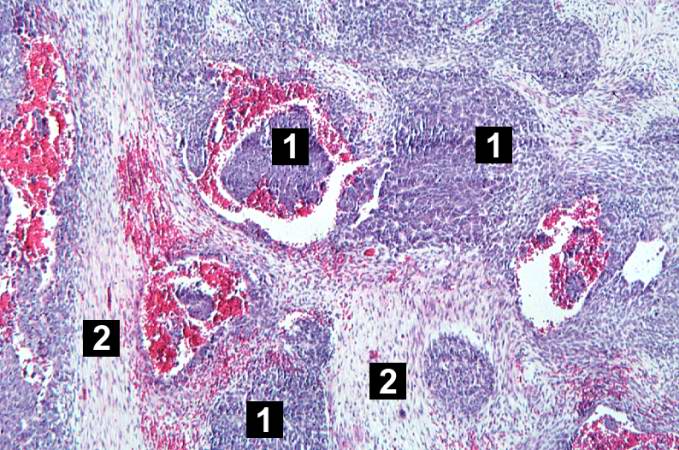 | 679 × 450 (91 KB) | Seung Park (talk | contribs) | This low-power photomicrograph of tumor shows the two cell types making up this neoplasm. The basophilic cellular component termed "blastema" (1) can be distinguished from less cellular eosinophilic areas with fibroblast-like cells (2). |
- You cannot overwrite this file.
File usage
The following page links to this file: