Difference between revisions of "File:IPLab13WT9.jpg"
Seung Park (talk | contribs) (This high-power photomicrograph demonstrates tubule formation within the blastema. Note the numerous mitotic figures (arrows).) |
(No difference)
|
Latest revision as of 05:56, 21 August 2013
This high-power photomicrograph demonstrates tubule formation within the blastema. Note the numerous mitotic figures (arrows).
File history
Click on a date/time to view the file as it appeared at that time.
| Date/Time | Thumbnail | Dimensions | User | Comment | |
|---|---|---|---|---|---|
| current | 05:56, 21 August 2013 | 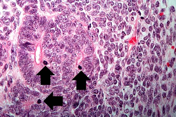 | 674 × 450 (81 KB) | Seung Park (talk | contribs) | This high-power photomicrograph demonstrates tubule formation within the blastema. Note the numerous mitotic figures (arrows). |
- You cannot overwrite this file.
File usage
The following page links to this file: