Difference between revisions of "File:IPLab1Prostate5.jpg"
Seung Park (talk | contribs) (This photomicrograph of prostatic epithelium demonstrates an in situ immunohistochemical technique that is used to identify the DNA fragments characteristic of apoptotic nuclei. This technique, terminal deoxynucleotidyl transferase-mediated dUTP-biotin...) |
(No difference)
|
Latest revision as of 03:42, 16 August 2013
This photomicrograph of prostatic epithelium demonstrates an in situ immunohistochemical technique that is used to identify the DNA fragments characteristic of apoptotic nuclei. This technique, terminal deoxynucleotidyl transferase-mediated dUTP-biotin nick end-labeling (TUNEL) is used to identify apoptotic cells (arrows) in histology sections.
File history
Click on a date/time to view the file as it appeared at that time.
| Date/Time | Thumbnail | Dimensions | User | Comment | |
|---|---|---|---|---|---|
| current | 03:42, 16 August 2013 | 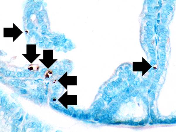 | 600 × 450 (35 KB) | Seung Park (talk | contribs) | This photomicrograph of prostatic epithelium demonstrates an in situ immunohistochemical technique that is used to identify the DNA fragments characteristic of apoptotic nuclei. This technique, terminal deoxynucleotidyl transferase-mediated dUTP-biotin... |
- You cannot overwrite this file.
File usage
The following page links to this file: