Difference between revisions of "File:IPLab2Hypertrophy1.jpg"
Seung Park (talk | contribs) (This is a gross photograph of a cross section of a normal human heart taken at autopsy (right) and the heart from this case, which demonstrates concentric hypertrophy of the left ventricular wall. Note the marked thickening of the left ventricular wall...) |
(No difference)
|
Latest revision as of 23:29, 18 August 2013
This is a gross photograph of a cross section of a normal human heart taken at autopsy (right) and the heart from this case, which demonstrates concentric hypertrophy of the left ventricular wall. Note the marked thickening of the left ventricular wall. There is also moderate thickening of the right ventricular wall.
Concentric hypertrophy is seen in pressure-overloaded hearts.
File history
Click on a date/time to view the file as it appeared at that time.
| Date/Time | Thumbnail | Dimensions | User | Comment | |
|---|---|---|---|---|---|
| current | 23:29, 18 August 2013 | 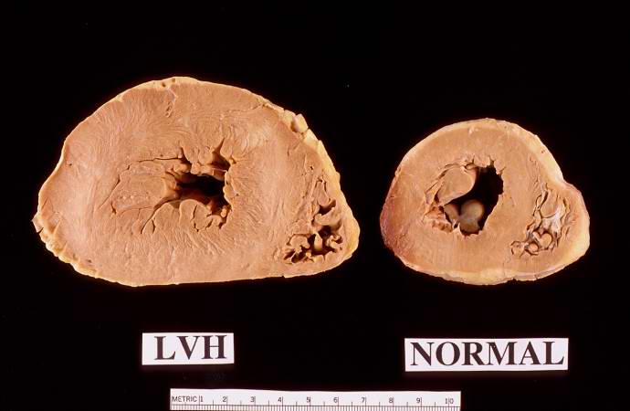 | 690 × 450 (30 KB) | Seung Park (talk | contribs) | This is a gross photograph of a cross section of a normal human heart taken at autopsy (right) and the heart from this case, which demonstrates concentric hypertrophy of the left ventricular wall. Note the marked thickening of the left ventricular wall... |
- You cannot overwrite this file.
File usage
The following page links to this file: