Difference between revisions of "File:IPLab2Hypertrophy6.jpg"
Seung Park (talk | contribs) (This gross photograph shows an example of normal physiologic hypertrophy. The organs shown are an open uterus (1), cervix (2) and vagina (3), both ovaries (4) and both kidneys (5) from a woman who died shortly after normal delivery from causes unrelate...) |
(No difference)
|
Latest revision as of 23:30, 18 August 2013
This gross photograph shows an example of normal physiologic hypertrophy. The organs shown are an open uterus (1), cervix (2) and vagina (3), both ovaries (4) and both kidneys (5) from a woman who died shortly after normal delivery from causes unrelated to childbirth. Note the marked thickening of the uterine wall due to smooth muscle cell hypertrophy.
File history
Click on a date/time to view the file as it appeared at that time.
| Date/Time | Thumbnail | Dimensions | User | Comment | |
|---|---|---|---|---|---|
| current | 23:30, 18 August 2013 | 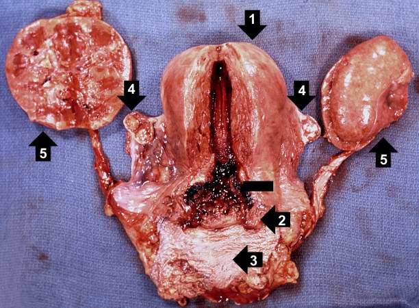 | 612 × 450 (63 KB) | Seung Park (talk | contribs) | This gross photograph shows an example of normal physiologic hypertrophy. The organs shown are an open uterus (1), cervix (2) and vagina (3), both ovaries (4) and both kidneys (5) from a woman who died shortly after normal delivery from causes unrelate... |
- You cannot overwrite this file.
File usage
The following page links to this file: