Difference between revisions of "File:IPLab3AcuteAppendicitis3.jpg"
Seung Park (talk | contribs) (This is a photomicrograph of an appendix exhibiting acute inflammation. Note that there are only remnants of mucosal tissue identifiable along the luminal border of this specimen. There is an extensive infiltration of leukocytes in this tissue which ca...) |
(No difference)
|
Latest revision as of 02:10, 19 August 2013
This is a photomicrograph of an appendix exhibiting acute inflammation. Note that there are only remnants of mucosal tissue identifiable along the luminal border of this specimen. There is an extensive infiltration of leukocytes in this tissue which cannot be specifically identified at this low power. Note that the surface is very roughened and has deposits of fibrin.
File history
Click on a date/time to view the file as it appeared at that time.
| Date/Time | Thumbnail | Dimensions | User | Comment | |
|---|---|---|---|---|---|
| current | 02:10, 19 August 2013 | 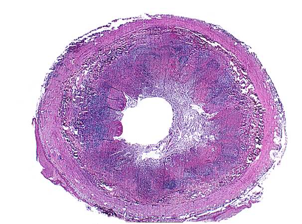 | 606 × 450 (64 KB) | Seung Park (talk | contribs) | This is a photomicrograph of an appendix exhibiting acute inflammation. Note that there are only remnants of mucosal tissue identifiable along the luminal border of this specimen. There is an extensive infiltration of leukocytes in this tissue which ca... |
- You cannot overwrite this file.
File usage
The following page links to this file: