Difference between revisions of "File:IPLab3Bronchopneumonia5.jpg"
Seung Park (talk | contribs) (This is a photomicrograph of lung tissue affected by bronchopneumonia. Note that the alveolar structure of this tissue, which is in the region of a terminal bronchiole (1), has been retained despite the massive infiltration of inflammatory cells. These...) |
(No difference)
|
Latest revision as of 03:23, 19 August 2013
This is a photomicrograph of lung tissue affected by bronchopneumonia. Note that the alveolar structure of this tissue, which is in the region of a terminal bronchiole (1), has been retained despite the massive infiltration of inflammatory cells. These inflammatory cells are primarily neutrophils.
File history
Click on a date/time to view the file as it appeared at that time.
| Date/Time | Thumbnail | Dimensions | User | Comment | |
|---|---|---|---|---|---|
| current | 03:23, 19 August 2013 | 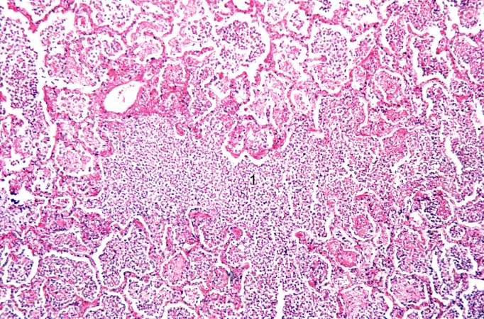 | 682 × 450 (113 KB) | Seung Park (talk | contribs) | This is a photomicrograph of lung tissue affected by bronchopneumonia. Note that the alveolar structure of this tissue, which is in the region of a terminal bronchiole (1), has been retained despite the massive infiltration of inflammatory cells. These... |
- You cannot overwrite this file.
File usage
The following page links to this file: