Difference between revisions of "File:IPLab3Sarcoidosis1.jpg"
Seung Park (talk | contribs) (This is a low-power photomicrograph of a lymph node. Note the rather pale-pink color of the tissue with dark-staining cells found in only a few scattered areas. These darker cells represent the original lymphocytes of this lymphoid organ.) |
(No difference)
|
Latest revision as of 03:29, 19 August 2013
This is a low-power photomicrograph of a lymph node. Note the rather pale-pink color of the tissue with dark-staining cells found in only a few scattered areas. These darker cells represent the original lymphocytes of this lymphoid organ.
File history
Click on a date/time to view the file as it appeared at that time.
| Date/Time | Thumbnail | Dimensions | User | Comment | |
|---|---|---|---|---|---|
| current | 03:29, 19 August 2013 | 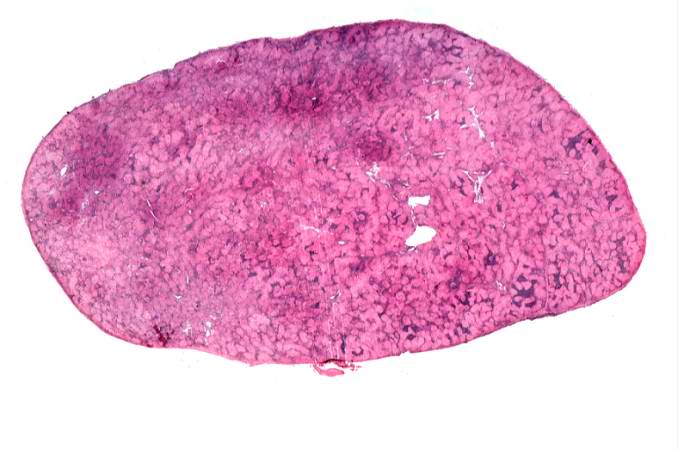 | 678 × 450 (40 KB) | Seung Park (talk | contribs) | This is a low-power photomicrograph of a lymph node. Note the rather pale-pink color of the tissue with dark-staining cells found in only a few scattered areas. These darker cells represent the original lymphocytes of this lymphoid organ. |
- You cannot overwrite this file.
File usage
The following page links to this file: