Difference between revisions of "File:IPLab3Tuberculosis4.jpg"
Seung Park (talk | contribs) (This is a high-power photomicrograph of a granuloma. Note the necrotic core on the right (1), epithelioid macrophages (2), and Langhans’ type giant cells (3) at the periphery of the granuloma. Note also the small lymphocytes characterized by their di...) |
(No difference)
|
Latest revision as of 03:39, 19 August 2013
This is a high-power photomicrograph of a granuloma. Note the necrotic core on the right (1), epithelioid macrophages (2), and Langhans’ type giant cells (3) at the periphery of the granuloma. Note also the small lymphocytes characterized by their distinctly blue-staining nuclei. Other cells in the tissue, in addition to the macrophages, include fibroblasts and occasional neutrophils.
File history
Click on a date/time to view the file as it appeared at that time.
| Date/Time | Thumbnail | Dimensions | User | Comment | |
|---|---|---|---|---|---|
| current | 03:39, 19 August 2013 | 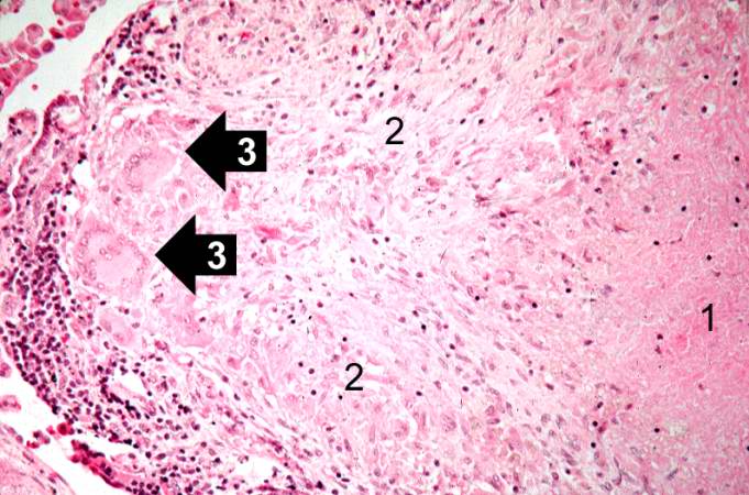 | 681 × 450 (66 KB) | Seung Park (talk | contribs) | This is a high-power photomicrograph of a granuloma. Note the necrotic core on the right (1), epithelioid macrophages (2), and Langhans’ type giant cells (3) at the periphery of the granuloma. Note also the small lymphocytes characterized by their di... |
- You cannot overwrite this file.
File usage
The following page links to this file: