Difference between revisions of "File:IPLab3ForeignBodyGranuloma3.jpg"
Seung Park (talk | contribs) (A higher-power photomicrograph of these granulomas reveals that they surround blood vessels (note the red blood cells within the lumen) (arrow).) |
(No difference)
|
Latest revision as of 03:47, 19 August 2013
A higher-power photomicrograph of these granulomas reveals that they surround blood vessels (note the red blood cells within the lumen) (arrow).
File history
Click on a date/time to view the file as it appeared at that time.
| Date/Time | Thumbnail | Dimensions | User | Comment | |
|---|---|---|---|---|---|
| current | 03:47, 19 August 2013 | 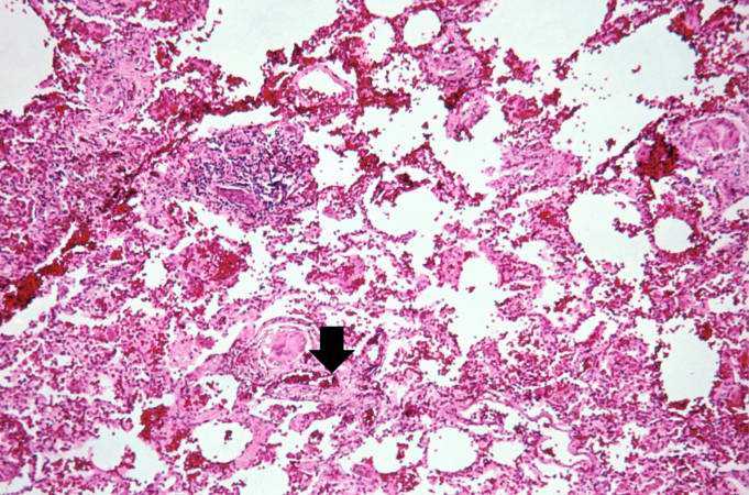 | 681 × 450 (89 KB) | Seung Park (talk | contribs) | A higher-power photomicrograph of these granulomas reveals that they surround blood vessels (note the red blood cells within the lumen) (arrow). |
- You cannot overwrite this file.
File usage
The following page links to this file: