Difference between revisions of "File:IPLab3ForeignBodyGranuloma4.jpg"
Seung Park (talk | contribs) (This is a photomicrograph of lung taken under light that is partially polarized to demonstrate the birefringent particles within the granulomas (1). Also, at this magnification one can better appreciate that these granulomas are adjacent to blood vesse...) |
(No difference)
|
Latest revision as of 03:48, 19 August 2013
This is a photomicrograph of lung taken under light that is partially polarized to demonstrate the birefringent particles within the granulomas (1). Also, at this magnification one can better appreciate that these granulomas are adjacent to blood vessels (2).
File history
Click on a date/time to view the file as it appeared at that time.
| Date/Time | Thumbnail | Dimensions | User | Comment | |
|---|---|---|---|---|---|
| current | 03:48, 19 August 2013 | 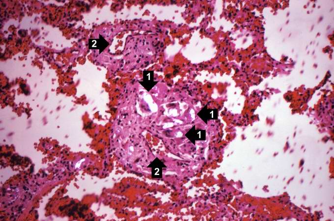 | 680 × 450 (62 KB) | Seung Park (talk | contribs) | This is a photomicrograph of lung taken under light that is partially polarized to demonstrate the birefringent particles within the granulomas (1). Also, at this magnification one can better appreciate that these granulomas are adjacent to blood vesse... |
- You cannot overwrite this file.
File usage
The following page links to this file: