Difference between revisions of "File:IPLab3FibrinousPericarditis3.jpg"
Seung Park (talk | contribs) (This low-power photomicrograph illustrates the dark-red-staining fibrin deposits on the inner surface (arrows). This pericardium is much thicker than normal and there are numerous inflammatory cells within the pericardial tissue.) |
(No difference)
|
Latest revision as of 04:10, 19 August 2013
This low-power photomicrograph illustrates the dark-red-staining fibrin deposits on the inner surface (arrows). This pericardium is much thicker than normal and there are numerous inflammatory cells within the pericardial tissue.
File history
Click on a date/time to view the file as it appeared at that time.
| Date/Time | Thumbnail | Dimensions | User | Comment | |
|---|---|---|---|---|---|
| current | 04:10, 19 August 2013 | 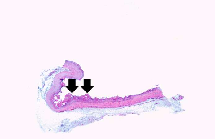 | 694 × 450 (13 KB) | Seung Park (talk | contribs) | This low-power photomicrograph illustrates the dark-red-staining fibrin deposits on the inner surface (arrows). This pericardium is much thicker than normal and there are numerous inflammatory cells within the pericardial tissue. |
- You cannot overwrite this file.
File usage
The following page links to this file: