Difference between revisions of "File:IPLab3FibrinousPericarditis5.jpg"
Seung Park (talk | contribs) (This high-power photomicrograph demonstrates fibrin (red amorphous material) on the surface of the pericardium (1). Note the reactive mesothelial cells on the surface of the pericardium (arrows) and the inflammatory cells within the pericardial tissue.) |
(No difference)
|
Latest revision as of 04:10, 19 August 2013
This high-power photomicrograph demonstrates fibrin (red amorphous material) on the surface of the pericardium (1). Note the reactive mesothelial cells on the surface of the pericardium (arrows) and the inflammatory cells within the pericardial tissue.
File history
Click on a date/time to view the file as it appeared at that time.
| Date/Time | Thumbnail | Dimensions | User | Comment | |
|---|---|---|---|---|---|
| current | 04:10, 19 August 2013 | 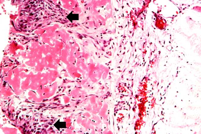 | 676 × 450 (64 KB) | Seung Park (talk | contribs) | This high-power photomicrograph demonstrates fibrin (red amorphous material) on the surface of the pericardium (1). Note the reactive mesothelial cells on the surface of the pericardium (arrows) and the inflammatory cells within the pericardial tissue. |
- You cannot overwrite this file.
File usage
The following page links to this file: