Difference between revisions of "File:IPLab3ChronicPepticUlcer2.jpg"
Seung Park (talk | contribs) (This is a gross photograph of the ulcer after it has been transected. The edge of the mucosa (1) is better appreciated in this image. Note the thick, fatty tissue (2) which makes up the base of this ulcer (3).) |
(No difference)
|
Latest revision as of 04:15, 19 August 2013
This is a gross photograph of the ulcer after it has been transected. The edge of the mucosa (1) is better appreciated in this image. Note the thick, fatty tissue (2) which makes up the base of this ulcer (3).
File history
Click on a date/time to view the file as it appeared at that time.
| Date/Time | Thumbnail | Dimensions | User | Comment | |
|---|---|---|---|---|---|
| current | 04:15, 19 August 2013 | 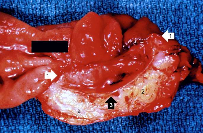 | 685 × 450 (61 KB) | Seung Park (talk | contribs) | This is a gross photograph of the ulcer after it has been transected. The edge of the mucosa (1) is better appreciated in this image. Note the thick, fatty tissue (2) which makes up the base of this ulcer (3). |
- You cannot overwrite this file.
File usage
The following page links to this file: