Difference between revisions of "File:IPLab3ChronicPepticUlcer4.jpg"
Seung Park (talk | contribs) (This is a photomicrograph of the margin of the ulcer. Note the intact epithelium on the right side of the section (1) and the ulcerated region without epithelium on the left (2). There are numerous inflammatory cells within this tissue.) |
(No difference)
|
Latest revision as of 04:15, 19 August 2013
This is a photomicrograph of the margin of the ulcer. Note the intact epithelium on the right side of the section (1) and the ulcerated region without epithelium on the left (2). There are numerous inflammatory cells within this tissue.
File history
Click on a date/time to view the file as it appeared at that time.
| Date/Time | Thumbnail | Dimensions | User | Comment | |
|---|---|---|---|---|---|
| current | 04:15, 19 August 2013 | 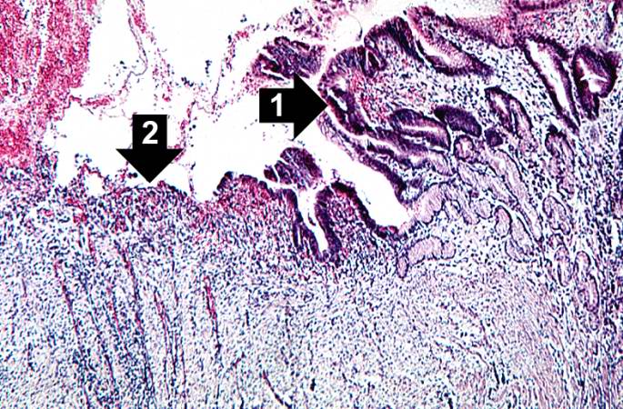 | 685 × 450 (104 KB) | Seung Park (talk | contribs) | This is a photomicrograph of the margin of the ulcer. Note the intact epithelium on the right side of the section (1) and the ulcerated region without epithelium on the left (2). There are numerous inflammatory cells within this tissue. |
- You cannot overwrite this file.
File usage
The following page links to this file: