Difference between revisions of "File:IPLab3ChronicPepticUlcer6.jpg"
Seung Park (talk | contribs) (This high-power photomicrograph of the ulcer base (arrows) demonstrates the lack of epithelium and the exuberant inflammatory response (1) consisting of primarily of fibrin (and adherent gastric secretions) and PMNs. The surface of the ulcer bed is cov...) |
(No difference)
|
Latest revision as of 04:16, 19 August 2013
This high-power photomicrograph of the ulcer base (arrows) demonstrates the lack of epithelium and the exuberant inflammatory response (1) consisting of primarily of fibrin (and adherent gastric secretions) and PMNs. The surface of the ulcer bed is covered with this fibrinopurulent exudate.
File history
Click on a date/time to view the file as it appeared at that time.
| Date/Time | Thumbnail | Dimensions | User | Comment | |
|---|---|---|---|---|---|
| current | 04:16, 19 August 2013 | 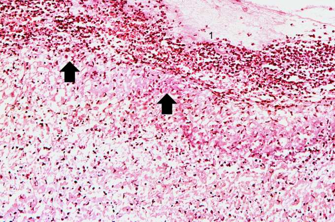 | 680 × 450 (92 KB) | Seung Park (talk | contribs) | This high-power photomicrograph of the ulcer base (arrows) demonstrates the lack of epithelium and the exuberant inflammatory response (1) consisting of primarily of fibrin (and adherent gastric secretions) and PMNs. The surface of the ulcer bed is cov... |
- You cannot overwrite this file.
File usage
The following page links to this file: