Difference between revisions of "File:IPLab3ChronicPepticUlcer7.jpg"
Seung Park (talk | contribs) (This high-power photomicrograph of the ulcer base demonstrates plump, activated fibroblasts and endothelial cells (arrows) within the granulation tissue that makes up the base of the ulcer. There are inflammatory cells (primarily lymphocytes) within th...) |
(No difference)
|
Latest revision as of 04:16, 19 August 2013
This high-power photomicrograph of the ulcer base demonstrates plump, activated fibroblasts and endothelial cells (arrows) within the granulation tissue that makes up the base of the ulcer. There are inflammatory cells (primarily lymphocytes) within this region as well.
File history
Click on a date/time to view the file as it appeared at that time.
| Date/Time | Thumbnail | Dimensions | User | Comment | |
|---|---|---|---|---|---|
| current | 04:16, 19 August 2013 | 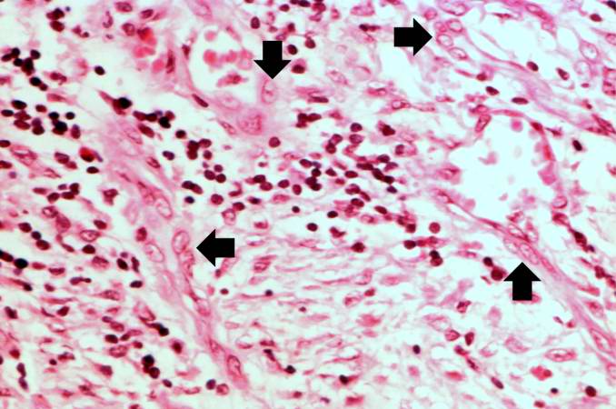 | 678 × 450 (53 KB) | Seung Park (talk | contribs) | This high-power photomicrograph of the ulcer base demonstrates plump, activated fibroblasts and endothelial cells (arrows) within the granulation tissue that makes up the base of the ulcer. There are inflammatory cells (primarily lymphocytes) within th... |
- You cannot overwrite this file.
File usage
The following page links to this file: