Difference between revisions of "File:IPLab3ChronicPepticUlcer11.jpg"
Seung Park (talk | contribs) (This is a trichrome-stained section of tissue demonstrating fibrous connective tissue scar formation (blue color) in this lesion. The surface of the ulcer is at the left-hand side of the image. There is a layer of inflammatory cells and RBCs on the sur...) |
(No difference)
|
Latest revision as of 04:17, 19 August 2013
This is a trichrome-stained section of tissue demonstrating fibrous connective tissue scar formation (blue color) in this lesion. The surface of the ulcer is at the left-hand side of the image. There is a layer of inflammatory cells and RBCs on the surface of the ulcer.
File history
Click on a date/time to view the file as it appeared at that time.
| Date/Time | Thumbnail | Dimensions | User | Comment | |
|---|---|---|---|---|---|
| current | 04:17, 19 August 2013 | 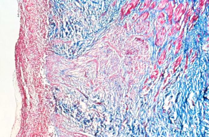 | 683 × 450 (73 KB) | Seung Park (talk | contribs) | This is a trichrome-stained section of tissue demonstrating fibrous connective tissue scar formation (blue color) in this lesion. The surface of the ulcer is at the left-hand side of the image. There is a layer of inflammatory cells and RBCs on the sur... |
- You cannot overwrite this file.
File usage
The following page links to this file: