Difference between revisions of "File:IPLab3BrainInfarction4.jpg"
Seung Park (talk | contribs) (This is a photomicrograph of the edge of the infarct. Note the numerous inflammatory cells in the brain parenchyma and adjacent to the remaining brain tissue (arrows).) |
(No difference)
|
Latest revision as of 04:22, 19 August 2013
This is a photomicrograph of the edge of the infarct. Note the numerous inflammatory cells in the brain parenchyma and adjacent to the remaining brain tissue (arrows).
File history
Click on a date/time to view the file as it appeared at that time.
| Date/Time | Thumbnail | Dimensions | User | Comment | |
|---|---|---|---|---|---|
| current | 04:22, 19 August 2013 | 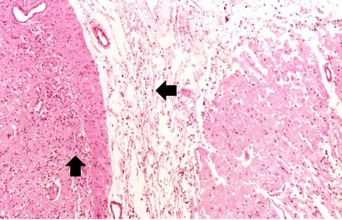 | 698 × 450 (68 KB) | Seung Park (talk | contribs) | This is a photomicrograph of the edge of the infarct. Note the numerous inflammatory cells in the brain parenchyma and adjacent to the remaining brain tissue (arrows). |
- You cannot overwrite this file.
File usage
The following page links to this file: