Difference between revisions of "File:IPLab3AcuteMyocardialInfarction4.jpg"
Seung Park (talk | contribs) (This is a higher-power photomicrograph of the edge of the infarct. The accumulation of inflammatory cells is on the left (1) and the infarcted tissue is on the right (2). Note that intact cells can be seen in the infarct but there are no nuclei.) |
(No difference)
|
Latest revision as of 04:30, 19 August 2013
This is a higher-power photomicrograph of the edge of the infarct. The accumulation of inflammatory cells is on the left (1) and the infarcted tissue is on the right (2). Note that intact cells can be seen in the infarct but there are no nuclei.
File history
Click on a date/time to view the file as it appeared at that time.
| Date/Time | Thumbnail | Dimensions | User | Comment | |
|---|---|---|---|---|---|
| current | 04:30, 19 August 2013 | 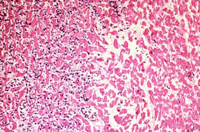 | 682 × 450 (79 KB) | Seung Park (talk | contribs) | This is a higher-power photomicrograph of the edge of the infarct. The accumulation of inflammatory cells is on the left (1) and the infarcted tissue is on the right (2). Note that intact cells can be seen in the infarct but there are no nuclei. |
- You cannot overwrite this file.
File usage
The following page links to this file: