Difference between revisions of "File:IPLab4MuralThrombus3.jpg"
Seung Park (talk | contribs) (This higher-power photomicrograph shows the border between the thrombus on the right (1) and the endocardium on the left (2). There is a line of inflammatory cells at this interface (arrow).) |
(No difference)
|
Latest revision as of 16:28, 19 August 2013
This higher-power photomicrograph shows the border between the thrombus on the right (1) and the endocardium on the left (2). There is a line of inflammatory cells at this interface (arrow).
A thrombus is a solid mass resulting from the aggregation of blood constituents within the vascular system.
File history
Click on a date/time to view the file as it appeared at that time.
| Date/Time | Thumbnail | Dimensions | User | Comment | |
|---|---|---|---|---|---|
| current | 16:28, 19 August 2013 | 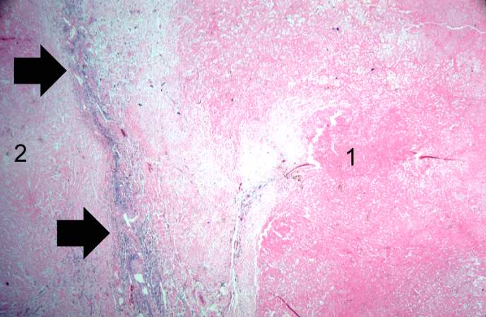 | 688 × 450 (53 KB) | Seung Park (talk | contribs) | This higher-power photomicrograph shows the border between the thrombus on the right (1) and the endocardium on the left (2). There is a line of inflammatory cells at this interface (arrow). |
- You cannot overwrite this file.
File usage
The following page links to this file: