Difference between revisions of "File:IPLab4AtheromatousEmboli1.jpg"
Seung Park (talk | contribs) (This is a gross photograph of the aorta from this patient opened lengthwise with the luminal surface visible. Note the rough surface with ulcerations and adherent thrombotic material. There is a mild dilation (aneurysm) at the distal aorta just at the ...) |
(No difference)
|
Latest revision as of 17:00, 19 August 2013
This is a gross photograph of the aorta from this patient opened lengthwise with the luminal surface visible. Note the rough surface with ulcerations and adherent thrombotic material. There is a mild dilation (aneurysm) at the distal aorta just at the bifurcation with an accumulation of thrombus.
A thrombus is a solid mass resulting from the aggregation of blood constituents within the vascular system.
File history
Click on a date/time to view the file as it appeared at that time.
| Date/Time | Thumbnail | Dimensions | User | Comment | |
|---|---|---|---|---|---|
| current | 17:00, 19 August 2013 | 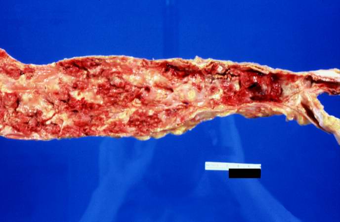 | 688 × 450 (31 KB) | Seung Park (talk | contribs) | This is a gross photograph of the aorta from this patient opened lengthwise with the luminal surface visible. Note the rough surface with ulcerations and adherent thrombotic material. There is a mild dilation (aneurysm) at the distal aorta just at the ... |
- You cannot overwrite this file.
File usage
The following page links to this file: