Difference between revisions of "File:IPLab6RA7.jpg"
Seung Park (talk | contribs) (This higher-power photomicrograph of the subcutaneous nodule again demonstrates the necrotic center and peripheral rim of macrophages, fibrocytes, and occasional lymphocytes. There are focal accumulations of hyaline material (fibrinoid material) within...) |
(No difference)
|
Latest revision as of 18:07, 19 August 2013
This higher-power photomicrograph of the subcutaneous nodule again demonstrates the necrotic center and peripheral rim of macrophages, fibrocytes, and occasional lymphocytes. There are focal accumulations of hyaline material (fibrinoid material) within the granuloma.
File history
Click on a date/time to view the file as it appeared at that time.
| Date/Time | Thumbnail | Dimensions | User | Comment | |
|---|---|---|---|---|---|
| current | 18:07, 19 August 2013 | 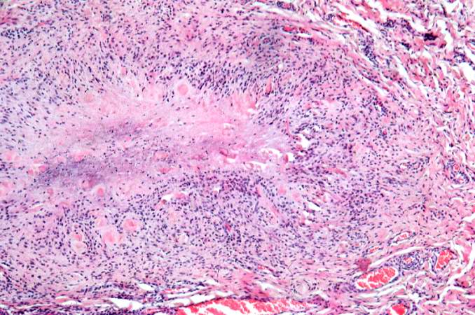 | 678 × 450 (82 KB) | Seung Park (talk | contribs) | This higher-power photomicrograph of the subcutaneous nodule again demonstrates the necrotic center and peripheral rim of macrophages, fibrocytes, and occasional lymphocytes. There are focal accumulations of hyaline material (fibrinoid material) within... |
- You cannot overwrite this file.
File usage
The following page links to this file: