Difference between revisions of "File:IPLab6RA8.jpg"
Seung Park (talk | contribs) (This higher-power photomicrograph of the tissue illustrates the palisading nuclei of the monocytes which are located around the periphery of the central necrotic region (1).) |
(No difference)
|
Latest revision as of 18:08, 19 August 2013
This higher-power photomicrograph of the tissue illustrates the palisading nuclei of the monocytes which are located around the periphery of the central necrotic region (1).
File history
Click on a date/time to view the file as it appeared at that time.
| Date/Time | Thumbnail | Dimensions | User | Comment | |
|---|---|---|---|---|---|
| current | 18:08, 19 August 2013 | 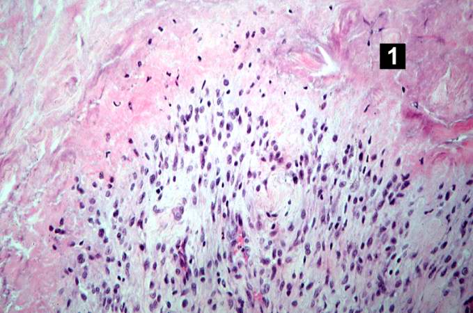 | 679 × 450 (59 KB) | Seung Park (talk | contribs) | This higher-power photomicrograph of the tissue illustrates the palisading nuclei of the monocytes which are located around the periphery of the central necrotic region (1). |
- You cannot overwrite this file.
File usage
The following page links to this file: