From Pathology Education Instructional Resource
Revision as of 18:09, 19 August 2013
Images
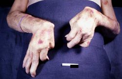
This is a gross photograph of the patient's hands at autopsy. Note the swollen joints and the deforming arthritis.
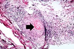
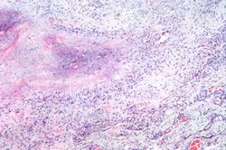
This is a medium-power photomicrograph of the joint capsule surrounding the metacarpal joints. Note the thickening of the capsule and the focal accumulation of inflammatory cells surrounding a central area of fibrinoid necrosis (arrow).
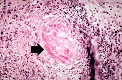
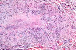
This is a high-power photomicrograph of the joint capsule with another granuloma surrounding a central area of fibrinoid necrosis (arrow).
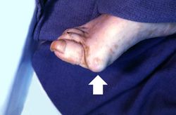
This is a gross photograph of the foot from this same patient. Note the subcutaneous nodule on the medial aspect of the foot (arrow).
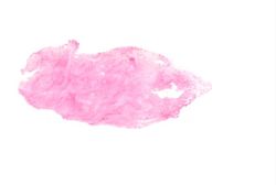
This is a low-power photomicrograph of the subcutaneous nodule from this patient.
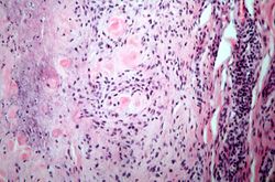
This higher-power photomicrograph of the subcutaneous nodule shows a granulomatous lesion with a necrotic center and a peripheral rim of macrophages, fibrocytes, and occasional lymphocytes. In the necrotic center of the granuloma there is some mineralization (basophilic material).
This higher-power photomicrograph of the subcutaneous nodule again demonstrates the necrotic center and peripheral rim of macrophages, fibrocytes, and occasional lymphocytes. There are focal accumulations of hyaline material (fibrinoid material) within the granuloma.
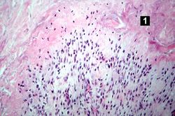
This higher-power photomicrograph of the tissue illustrates the palisading nuclei of the monocytes which are located around the periphery of the central necrotic region (1).
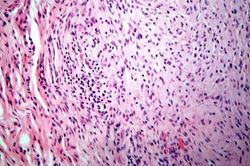
This is a high-power photomicrograph of the mononuclear cells which surround the central area of necrosis. The focal accumulations of fibrinoid material are clearly visible. Lymphocytes are present in the extreme right of this image.
This is a high-power photomicrograph of another region with macrophages (right), fibrocytes (left), and occasional lymphocytes throughout the lesion.
IPLab:Lab 6
