Difference between revisions of "File:IPLab5Downs1.jpg"
Seung Park (talk | contribs) (This is a photomicrograph of cells obtained by amniocentesis that were stained using FISH. The cell in panel 1 was stained with markers specific for the X and Y-chromosomes. The cell in panel 2 was stained with a marker specific for chromosome 18. The ...) |
(No difference)
|
Latest revision as of 18:27, 19 August 2013
This is a photomicrograph of cells obtained by amniocentesis that were stained using FISH. The cell in panel 1 was stained with markers specific for the X and Y-chromosomes. The cell in panel 2 was stained with a marker specific for chromosome 18. The cell in the center was stained with markers for chromosomes 13 and 21. Note that there are three copies of chromosome 21.
Amniocentesis is a procedure in which a needle is inserted transabdominally through the uterus, into the amniotic sac, and amniotic fluid is withdrawn.
File history
Click on a date/time to view the file as it appeared at that time.
| Date/Time | Thumbnail | Dimensions | User | Comment | |
|---|---|---|---|---|---|
| current | 18:27, 19 August 2013 | 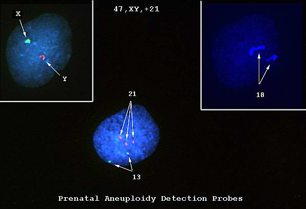 | 596 × 407 (18 KB) | Seung Park (talk | contribs) | This is a photomicrograph of cells obtained by amniocentesis that were stained using FISH. The cell in panel 1 was stained with markers specific for the X and Y-chromosomes. The cell in panel 2 was stained with a marker specific for chromosome 18. The ... |
- You cannot overwrite this file.
File usage
The following page links to this file: