Difference between revisions of "IPLab:Lab 5:Trisomy 21"
Seung Park (talk | contribs) (Created page with "== Images == <gallery heights="250px" widths="250px"> File:IPLab5Downs1.jpg|This is a photomicrograph of cells obtained by amniocentesis that were stained using FISH. The cell...") |
(No difference)
|
Revision as of 18:29, 19 August 2013
Images[edit]
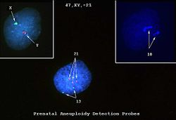
This is a photomicrograph of cells obtained by amniocentesis that were stained using FISH. The cell in panel 1 was stained with markers specific for the X and Y-chromosomes. The cell in panel 2 was stained with a marker specific for chromosome 18. The cell in the center was stained with markers for chromosomes 13 and 21. Note that there are three copies of chromosome 21.
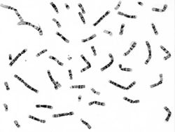
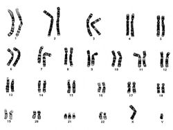
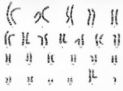
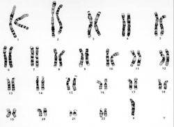
These cells, obtained by amniocentesis, were cultured and then arrested in metaphase. Nuclei from these cells were isolated and stained to demonstrate the banding pattern of each chromosome. This photograph shows a "chromosome spread." Each chromosome is identified and lined up to give a karyotype (next page).
| |||||
Amniocentesis is a procedure in which a needle is inserted transabdominally through the uterus, into the amniotic sac, and amniotic fluid is withdrawn.
