Difference between revisions of "File:IPLab6Amyloid9.jpg"
Seung Park (talk | contribs) (This is a gross photograph of kidney from this case. Note the pale yellow material within the cortex (arrows). This is indicative of amyloid within the cortex and the glomeruli. Also note that there are multiple red spots in the cortex. These represent...) |
(No difference)
|
Latest revision as of 21:36, 20 August 2013
This is a gross photograph of kidney from this case. Note the pale yellow material within the cortex (arrows). This is indicative of amyloid within the cortex and the glomeruli. Also note that there are multiple red spots in the cortex. These represent congested glomeruli due to the vascular compromise produced by the amyloid.
File history
Click on a date/time to view the file as it appeared at that time.
| Date/Time | Thumbnail | Dimensions | User | Comment | |
|---|---|---|---|---|---|
| current | 21:36, 20 August 2013 | 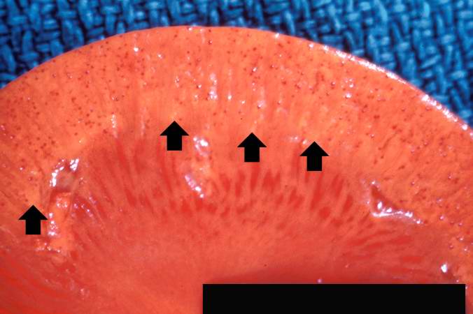 | 676 × 450 (34 KB) | Seung Park (talk | contribs) | This is a gross photograph of kidney from this case. Note the pale yellow material within the cortex (arrows). This is indicative of amyloid within the cortex and the glomeruli. Also note that there are multiple red spots in the cortex. These represent... |
- You cannot overwrite this file.
File usage
The following page links to this file: