Difference between revisions of "File:IPLab6Amyloid11.jpg"
Seung Park (talk | contribs) (This photomicrograph of the tongue demonstrates extensive amyloid deposits (1) separating the skeletal muscle fibers of the tongue. In many cases the amyloid encircles the muscle fibers (2) and these muscle fibers are atrophied.) |
(No difference)
|
Latest revision as of 21:36, 20 August 2013
This photomicrograph of the tongue demonstrates extensive amyloid deposits (1) separating the skeletal muscle fibers of the tongue. In many cases the amyloid encircles the muscle fibers (2) and these muscle fibers are atrophied.
File history
Click on a date/time to view the file as it appeared at that time.
| Date/Time | Thumbnail | Dimensions | User | Comment | |
|---|---|---|---|---|---|
| current | 21:36, 20 August 2013 | 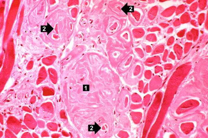 | 681 × 450 (58 KB) | Seung Park (talk | contribs) | This photomicrograph of the tongue demonstrates extensive amyloid deposits (1) separating the skeletal muscle fibers of the tongue. In many cases the amyloid encircles the muscle fibers (2) and these muscle fibers are atrophied. |
- You cannot overwrite this file.
File usage
The following page links to this file: