Difference between revisions of "File:IPLab6ChronicRejection4.jpg"
Seung Park (talk | contribs) (This is another area of renal cortex similar to the previous image. Note the fibrosis (1) and loss of renal tubules throughout this section. Also note the focus of inflammatory cells (2) indicating that despite the chromic nature of this lesion, there ...) |
(No difference)
|
Latest revision as of 21:49, 20 August 2013
This is another area of renal cortex similar to the previous image. Note the fibrosis (1) and loss of renal tubules throughout this section. Also note the focus of inflammatory cells (2) indicating that despite the chromic nature of this lesion, there is still ongoing active rejection and renal damage.
File history
Click on a date/time to view the file as it appeared at that time.
| Date/Time | Thumbnail | Dimensions | User | Comment | |
|---|---|---|---|---|---|
| current | 21:49, 20 August 2013 | 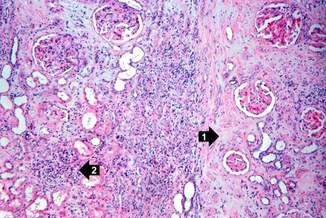 | 673 × 450 (84 KB) | Seung Park (talk | contribs) | This is another area of renal cortex similar to the previous image. Note the fibrosis (1) and loss of renal tubules throughout this section. Also note the focus of inflammatory cells (2) indicating that despite the chromic nature of this lesion, there ... |
- You cannot overwrite this file.
File usage
The following page links to this file: