Difference between revisions of "File:IPLab6ChronicRejection12.jpg"
Seung Park (talk | contribs) (Photomicrograph from another region of previous image. Note the cellular infiltrate around a small blood vessel (right) and neutrophils within renal tubules (arrow).) |
(No difference)
|
Latest revision as of 21:51, 20 August 2013
Photomicrograph from another region of previous image. Note the cellular infiltrate around a small blood vessel (right) and neutrophils within renal tubules (arrow).
An infiltrate is an accumulation of cells in the lung parenchyma--this is a sign of pneumonia.
File history
Click on a date/time to view the file as it appeared at that time.
| Date/Time | Thumbnail | Dimensions | User | Comment | |
|---|---|---|---|---|---|
| current | 21:51, 20 August 2013 | 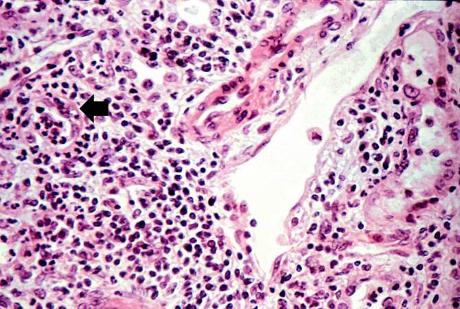 | 670 × 450 (72 KB) | Seung Park (talk | contribs) | Photomicrograph from another region of previous image. Note the cellular infiltrate around a small blood vessel (right) and neutrophils within renal tubules (arrow). |
- You cannot overwrite this file.
File usage
The following page links to this file: