Difference between revisions of "File:IPLab6AcuteRejection4.jpg"
Seung Park (talk | contribs) (This is a higher-power photomicrograph demonstrating the cellular infiltrates within this kidney section. Note that in addition to the diffuse cellularity, the focal accumulations of cells appear to be focused around blood vessels.) |
(No difference)
|
Latest revision as of 21:57, 20 August 2013
This is a higher-power photomicrograph demonstrating the cellular infiltrates within this kidney section. Note that in addition to the diffuse cellularity, the focal accumulations of cells appear to be focused around blood vessels.
An infiltrate is an accumulation of cells in the lung parenchyma--this is a sign of pneumonia.
File history
Click on a date/time to view the file as it appeared at that time.
| Date/Time | Thumbnail | Dimensions | User | Comment | |
|---|---|---|---|---|---|
| current | 21:57, 20 August 2013 | 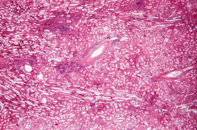 | 684 × 450 (78 KB) | Seung Park (talk | contribs) | This is a higher-power photomicrograph demonstrating the cellular infiltrates within this kidney section. Note that in addition to the diffuse cellularity, the focal accumulations of cells appear to be focused around blood vessels. |
- You cannot overwrite this file.
File usage
The following page links to this file: