Difference between revisions of "File:IPLab7LipSCC3.jpg"
Seung Park (talk | contribs) (This photomicrograph shows a large area of ulceration (arrow) with underlying congestion and hemorrhage. The area of ulceration is adjacent to an area of tumor infiltration.) |
(No difference)
|
Latest revision as of 01:32, 21 August 2013
This photomicrograph shows a large area of ulceration (arrow) with underlying congestion and hemorrhage. The area of ulceration is adjacent to an area of tumor infiltration.
File history
Click on a date/time to view the file as it appeared at that time.
| Date/Time | Thumbnail | Dimensions | User | Comment | |
|---|---|---|---|---|---|
| current | 01:32, 21 August 2013 | 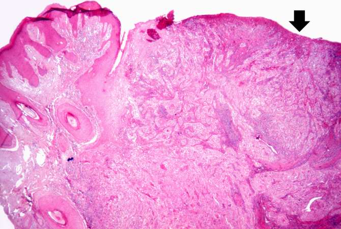 | 670 × 450 (57 KB) | Seung Park (talk | contribs) | This photomicrograph shows a large area of ulceration (arrow) with underlying congestion and hemorrhage. The area of ulceration is adjacent to an area of tumor infiltration. |
- You cannot overwrite this file.
File usage
The following page links to this file: