Difference between revisions of "File:IPLab7Metastatic8.jpg"
Seung Park (talk | contribs) (This is a high-power photomicrograph of the edge of the tumor nodule in the lung. The tumor cells are infiltrating into the lung parenchyma (1). Even at this power you can see the glandular formation of this adenocarcinoma. There is a large area of nec...) |
(No difference)
|
Latest revision as of 01:47, 21 August 2013
This is a high-power photomicrograph of the edge of the tumor nodule in the lung. The tumor cells are infiltrating into the lung parenchyma (1). Even at this power you can see the glandular formation of this adenocarcinoma. There is a large area of necrosis in the center of the tumor (2).
File history
Click on a date/time to view the file as it appeared at that time.
| Date/Time | Thumbnail | Dimensions | User | Comment | |
|---|---|---|---|---|---|
| current | 01:47, 21 August 2013 | 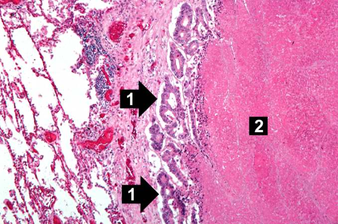 | 679 × 450 (71 KB) | Seung Park (talk | contribs) | This is a high-power photomicrograph of the edge of the tumor nodule in the lung. The tumor cells are infiltrating into the lung parenchyma (1). Even at this power you can see the glandular formation of this adenocarcinoma. There is a large area of nec... |
- You cannot overwrite this file.
File usage
The following page links to this file: