From Pathology Education Instructional Resource
Revision as of 01:47, 21 August 2013
Images
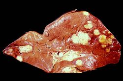
This gross photograph of the liver from this case demonstrates multiple, variably-sized pale/white-tan nodules scattered throughout the liver.
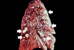
This gross photograph of the lung from this case also demonstrates multiple, variably sized pale/white-tan nodules scattered throughout the lung.
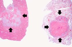
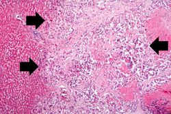
These are low-power photomicrographs of a section of liver (left) and lung (right) containing tumor nodules (arrows).
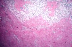
This is a photomicrograph taken at the interface between the tumor (top) and the normal liver parenchyma (bottom).
This is a higher-power photomicrograph showing how the tumor cells (arrows) have infiltrated into the liver parenchyma.
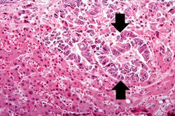
This is a high-power photomicrograph of tumor cells that are forming glands (arrows).
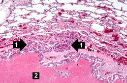
This is a photomicrograph of a tumor nodule in the lung. The tumor cells are infiltrating into the lung parenchyma (1). There is a large area of necrosis in the center of the tumor (2).
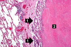
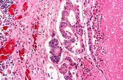
This is a high-power photomicrograph of the edge of the tumor nodule in the lung. The tumor cells are infiltrating into the lung parenchyma (1). Even at this power you can see the glandular formation of this adenocarcinoma. There is a large area of necrosis in the center of the tumor (2).
This is a high-power photomicrograph of the edge of the tumor nodule in the lung. The tumor cells area growing in a glandular pattern. The area of necrosis is evident at the right side of the image.
