Difference between revisions of "File:IPLab7Melanoma3.jpg"
Seung Park (talk | contribs) (This is a low-power photomicrograph of lymph node that is almost completely replaced/filled with tumor. This lymph node has a capsule (1) and some remaining lymphocytes (2) but the remainder of the node is replaced by tumor cells.) |
(No difference)
|
Latest revision as of 01:55, 21 August 2013
This is a low-power photomicrograph of lymph node that is almost completely replaced/filled with tumor. This lymph node has a capsule (1) and some remaining lymphocytes (2) but the remainder of the node is replaced by tumor cells.
File history
Click on a date/time to view the file as it appeared at that time.
| Date/Time | Thumbnail | Dimensions | User | Comment | |
|---|---|---|---|---|---|
| current | 01:55, 21 August 2013 | 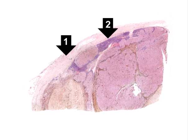 | 604 × 450 (21 KB) | Seung Park (talk | contribs) | This is a low-power photomicrograph of lymph node that is almost completely replaced/filled with tumor. This lymph node has a capsule (1) and some remaining lymphocytes (2) but the remainder of the node is replaced by tumor cells. |
- You cannot overwrite this file.
File usage
The following page links to this file: