Difference between revisions of "File:IPLab7Melanoma7.jpg"
Seung Park (talk | contribs) (This is a high-power photomicrograph of the main tumor mass with the cells growing as poorly formed nests and sheets of cells. There is little if any pigment in this section.) |
(No difference)
|
Latest revision as of 01:56, 21 August 2013
This is a high-power photomicrograph of the main tumor mass with the cells growing as poorly formed nests and sheets of cells. There is little if any pigment in this section.
File history
Click on a date/time to view the file as it appeared at that time.
| Date/Time | Thumbnail | Dimensions | User | Comment | |
|---|---|---|---|---|---|
| current | 01:56, 21 August 2013 | 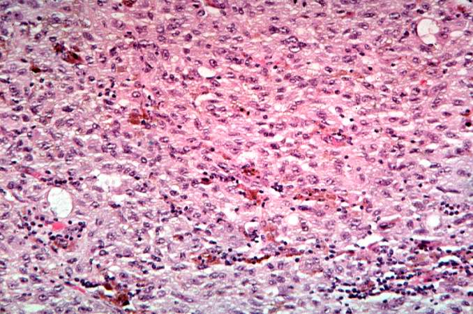 | 678 × 450 (81 KB) | Seung Park (talk | contribs) | This is a high-power photomicrograph of the main tumor mass with the cells growing as poorly formed nests and sheets of cells. There is little if any pigment in this section. |
- You cannot overwrite this file.
File usage
The following page links to this file: